Improving 18F-FDG PET Quantification Through a Spatial Normalization Method
- 작성자
- 관리자
- 등록일
- 2025-03-06
Background: Quantification of 18F-FDG PET images is useful for accurate diagnosis and evaluation of various brain diseases, including brain tumors, epilepsy, dementia, and Parkinson disease. However, accurate quantification of 18F-FDG PET images requires matched 3-dimensional T1 MRI scans of the same individuals to provide detailed information on brain anatomy. In this paper, we propose a transfer learning approach to adapt a pretrained deep neural network model from amyloid PET to spatially normalize 18F-FDG PET images without the need for 3-dimensional MRI.
Methods: The proposed method is
based on a deep learning model for automatic spatial normalization of 18F-FDG
brain PET images, which was developed by fine-tuning a pretrained model for
amyloid PET using only 103 18F-FDG PET and MR images. After training, the
algorithm was tested on 65 internal and 78 external test sets. All T1 MR
images with a 1-mm isotropic voxel size were processed with FreeSurfer software
to provide cortical segmentation maps used to extract a ground-truth regional
SUV ratio using cerebellar gray matter as a reference region. These values were
compared with those from spatial normalization-based quantification methods
using the proposed method and statistical parametric mapping software.
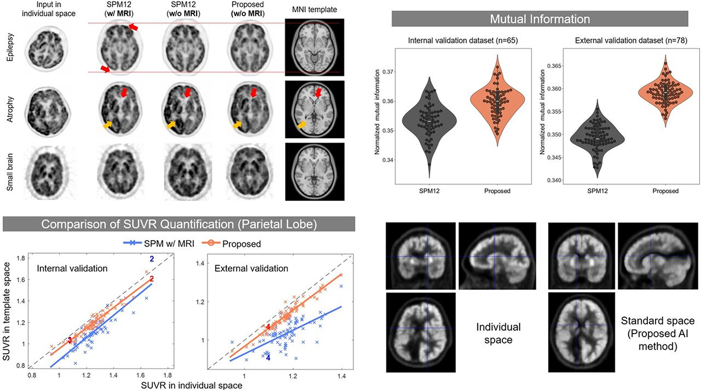
Results: The proposed method showed superior spatial normalization compared with statistical parametric mapping, as evidenced by increased normalized mutual information and better size and shape matching in PET images. Quantitative evaluation revealed a consistently higher SUV ratio correlation and intraclass correlation coefficients for the proposed method across various brain regions in both internal and external datasets. The remarkably good correlation and intraclass correlation coefficient values of the proposed method for the external dataset are noteworthy, considering the dataset’s different ethnic distribution and the use of different PET scanners and image reconstruction algorithms.
Conclusion: This study
successfully applied transfer learning to a deep neural network for 18F-FDG
PET spatial normalization, demonstrating its resource efficiency and improved
performance. This highlights the efficacy of transfer learning, which requires
a smaller number of datasets than does the original network training, thus
increasing the potential for broader use of deep learning–based brain PET
spatial normalization techniques for various clinical and research
radiotracers.
Keywords: brain PET, quantification,
spatial normalization, glucose metabolism
Journal
of Nuclear Medicine August 2024, jnumed.123.267360; DOI:
https://doi.org/10.2967/jnumed.123.267360
Link: https://jnm.snmjournals.org/content/early/2024/08/29/jnumed.123.267360